Overview
MRI stands for magnetic resonance imaging. It uses radio waves, powerful magnets and a computer to take detailed pictures inside your body.
An MRI is a noninvasive way for doctors to look at the inside of your body. Noninvasive means that it doesn’t require your doctor to cut your skin or put any instruments inside your body.
Types of MRI Scanners
MRI machines are shaped like a long tube that is open at both ends. When you lie inside the machine, it takes cross-sectional images of your body. The images are like slices from a loaf of bread.
We have five scanners at the medical center. The magnets include three, 1.5 Tesla scanners and two, 3 Tesla scanners. The type of your exam and results of your screening will to help us decide which scanner to use.
Brain and Spinal Cord Imaging
Doctors use MRI to take pictures of the brain and spinal cord. It can help diagnose:
- Brain aneurysms (a bulge in a blood vessel)
- Brain injuries caused by trauma
- Eye and ear disorders
- Multiple sclerosis
- Spinal cord problems
- Strokes
- Tumors
Images of the Heart and Blood Vessels
When doctors use this test to look at the blood vessels and the heart, they can find things like:
- How well the heart is working
- The heart’s thickness
- If it’s been damaged by heart disease or heart attacks
- Problems in the aorta (the main blood vessel)
- Blockages or swelling in the blood vessels
MRI of Other Organs
This exam can be used to check for tumors or conditions of many of the body’s organs. These include:
- Kidneys
- Liver and bile ducts
- Ovaries
- Pancreas
- Prostate
- Spleen
- Uterus
Bone and Joint MRI
When doctors want to look inside the bones and joints, and MRI can help. They can see:
- Bone infections
- Torn cartilage, ligaments or other joint abnormalities
- Problems with the spine
- Bone and soft tissue tumors
Breast MRI
Doctors use MRI with mammograms to find breast cancer. Learn more about breast MRI
Is MRI Safe?
Yes, it is safe to get an MRI when proper rules are followed. We take extra safety measures before your exam with a thorough screening process.
MRI Screening Process
Our goal is, first and foremost, to protect your safety. The strong magnetic field used for MRI will pull on any objects in your body with iron in them.
As a result, we have a two-part screening process. The first part takes place before the exam. The second part happens the day of the exam.
Before Your Exam – Medical History
Before your exam, we’ll ask you to fill out a form, telling us about your medical history. You may feel like the same questions are being asked more than once, but that is part of our screening process for your safety.
You can fill out this form one of three ways:
- A phone call before the exam
- At home before your exam
- The day of your exam
The form asks a lot of questions about your medical history, so completing it early will save you time in registration on the day of your appointment.
This screening form will help keep you safe by asking you to share any information about your health and medical history. We will focus on anything you have inside or outside your body you were not born with.
This includes any:
- Implants
- Pacemakers
- Clips
- IVC Filters
- Other devices checked on the first page of the screening form
If you have any medical paperwork about these objects, also called an implant record, please bring it to your appointment. It is important for us to have it so we can ensure your safety.
Before the MRI, our technologists will review the model numbers of each device to make sure you’ll be safe while you’re in the machine. This will also tell us which machine we’ll use for your exam.
If you do have a pacemaker, stimulator or other electronic device, the technologist may ask a representative from the company to be present during your scan. This will help ensure the safety of the device during your scan.
The Day of Your Exam – Metal/Magnetic Detection
On the day of your exam, you will go through a metal/magnetic detector. That helps us make sure there is no metal in or on your body that can react to the extremely strong magnets in the scanners.
During this part of the process, you’ll walk past a metal detector mounted on the wall. We may also scan you with a handheld device like the ones you see in airport security.
If you have had surgery to take out any metal, bullet or shrapnel from your eyes, or you have ever worked with metal, or have certain implants, we may send you to our X-ray department before your test. This is another way we can help keep you safe during your MRI.
What to Wear
Many types of fabric and shoes contain metal. So, for your safety we will provide you with a hospital gown, pants and socks for you to change into before your exam. Please take off, or leave any jewelry, like earrings, necklaces or bracelets at home. We’ll give you a locker with a lock to store everything safely. The University of Tennessee Medical Center is not responsible for lost or stolen items.
What to Expect During Your Exam
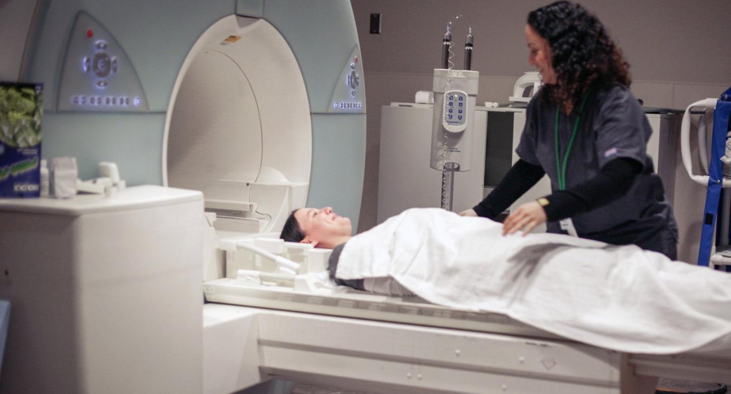
The MRI machine looks like a long narrow tube that’s open on both sides. In the exam room, we will help you lie down on a padded, moveable table and make you as comfortable as possible for your exam.
Your technologist will be outside the room, but you will have a bulb to squeeze to communicate via microphone, if needed.
An MRI doesn’t hurt, but some patients find it hard to stay still during the exam. You may also hear a loud knocking noise during the exam, which is normal – it’s just the machine doing its job.
To protect your hearing, we will give you earplugs and headphones. We will also let you choose music to listen to during the MRI.
Your ordering physician may request MRI contrast (dye) material for your exam, based on the reason you are having the test. You may be given a drink with the dye in it, or it may be injected into your veins through an intravenous (IV) line.
An exam can take anywhere from 15 minutes to several hours, depending on the kind of exam you’re having. It is important for you to stay still during the exam. That’s because any movement may blur the images. That means we have to re-take the images, and this could make your exam last longer.
Please tell your ordering physician if you are claustrophobic, or are afraid of enclosed spaces. They can decide if you need medication to help you relax for your exam.
What to Expect After Your Exam
Once the exam is finished, the technologist will help you to the dressing room to change clothes and collect your valuables. If you haven’t been given a sedative, you’ll be able to go back to your normal routine right away.
When Will I Get My Results?
The University of Tennessee Medical Center’s Radiology Department is committed to giving you the best care. Your ordering physician will receive the report from your exam within three business days. They will notify you of your results and plan your future care.