What is a Breast Ultrasound?
A breast ultrasound is a safe, painless and noninvasive medical test. It helps doctors diagnose and treat medical conditions of the breast.
Ultrasound can be used as a screening tool for women who:
- Are at high risk for breast cancer and unable to undergo an MRI examination.
- Are pregnant or should not be exposed to x-rays, which is necessary for a mammogram.
How Is a Breast Ultrasound Performed?
During your test, the sonographer — the person doing the ultrasounds — will position you on an examination table.
After you are positioned on the table, the radiologist or sonographer will apply a warm water-based gel to the breast. The gel helps make secure contact with the body. It also gets rid of air pockets between the probe and the skin that can block the sound waves from passing into your body.
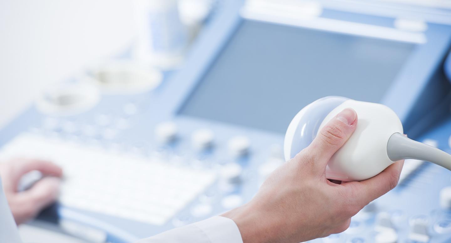
The sonographer will then run a probe, called a tranducer, back and forth over the area of interest. This makes images of the inside of the breast.
Doppler ultrasound is a special ultrasound technique that evaluates blood flow through a blood vessel. During a breast ultrasound, the sonographer may use Doppler to check the blood flow in any breast mass. Sometimes this gives your doctor more information about the cause of the mass.
Patients usually don’t feel any discomfort during the test. However, if the sonographer scans an area of tenderness, you may feel pressure or minor pain from the probe.
Once the imaging is complete, the clear ultrasound gel will be wiped off your skin. The ultrasound gel does not stain or discolor clothing.
Who Interprets the Results and How Do I Get Them?
A radiologist is a physician specifically trained to supervise and interpret radiology examinations. Your radiologist will analyze the images and send a signed report to the doctor or other health care provider who ordered the test. Your provider (not the sonographer or radiologist) will share the results with you.
If you need a follow-up examination, your doctor will explain why.
What Are the Benefits Versus Risks?
Like most medical tests, ultrasounds carry benefits and risks. You can read about the general benefits and risks on the ultrasound page. This list covers the benefits and risks related just to breast ultrasounds.
Benefits
Ultrasound imaging can help detect lesions in women with dense breasts.
- Ultrasound may help detect and classify a breast lesion that cannot be interpreted adequately through mammography alone.
- Using ultrasound, physicians are able to determine that many areas of clinical concern are due to normal tissue, such as fat lobules, or benign cysts. For most women 30 or older, a mammogram will be used together with ultrasound. For women under 30, ultrasound alone is often sufficient to determine whether an area of concern needs a biopsy or not.
Risks
- Interpretation of a breast ultrasound examination may lead to additional procedures such as follow-up ultrasound and/or aspiration or biopsy. Many of the areas thought to be of concern on ultrasound turn out to be non-cancerous.


